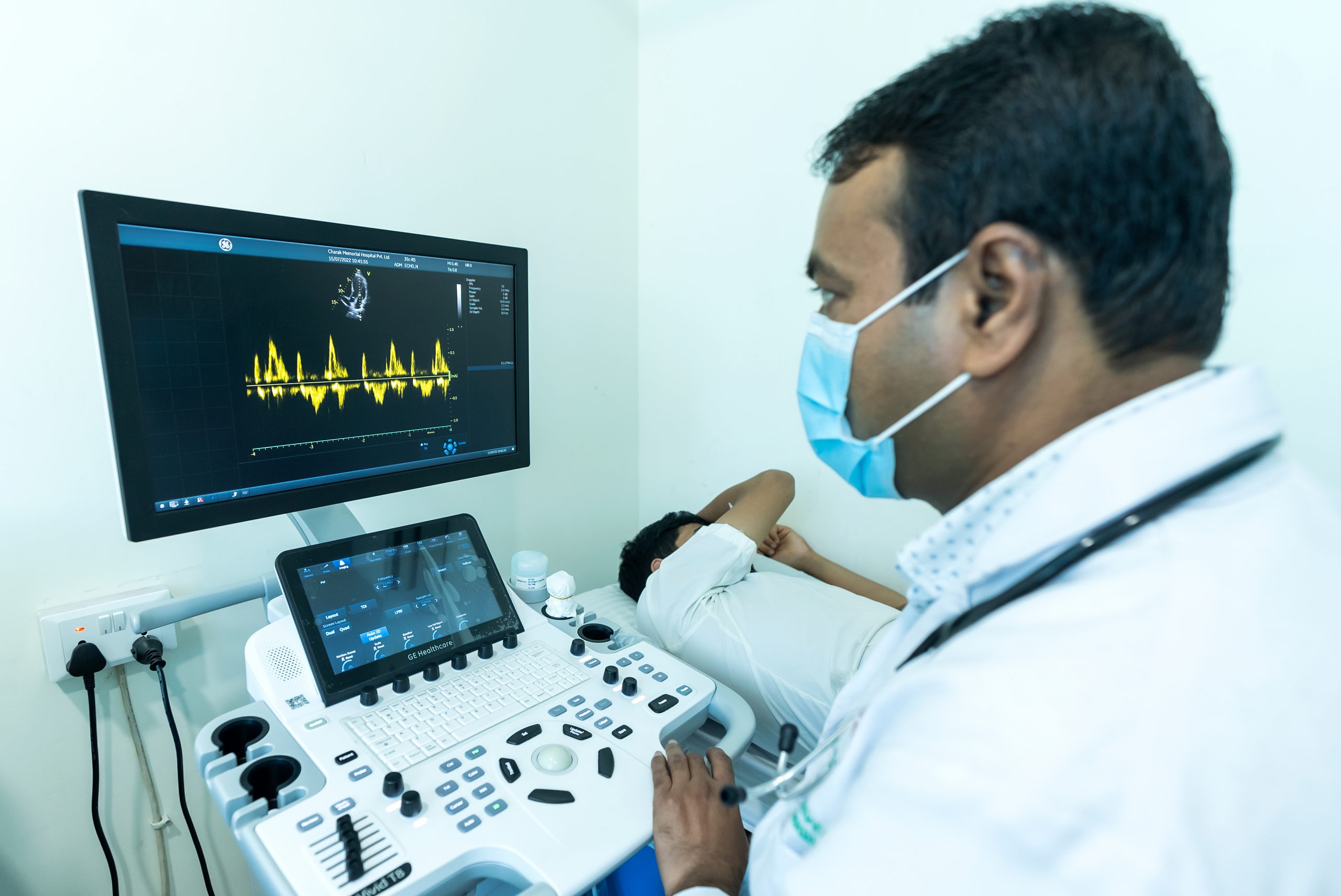
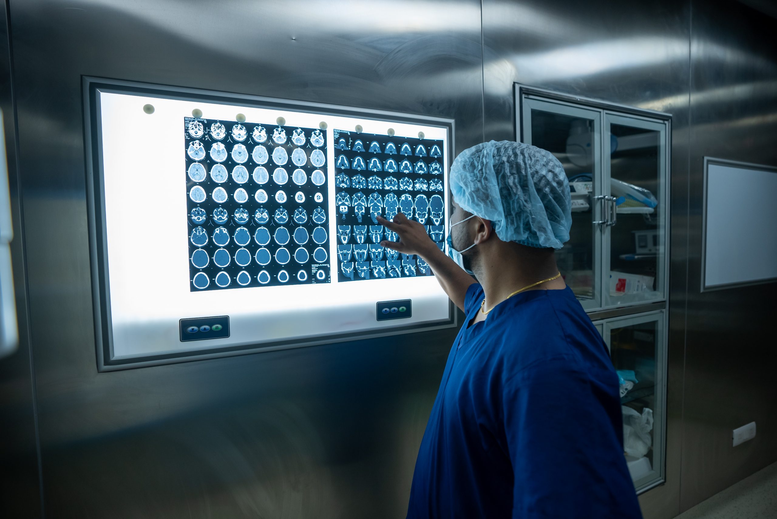
Radiology uses medical imaging technology to diagnose and treat diseases. Basically, radiology involves imaging the inside of the human body. It uses different imaging modalities like electromagnetic radiation, ultrasonography, magnetic resonance, fluoroscopy, nuclear medicine, positron emission tomography, etc to give the result.
There are following branches of radiology:
ECG
Electrocardiogram is used to check heart’s rhythm and electrical activity. Sensors attached to the skin are used to detect the electrical signals produced by heart each time it beats. Change in the ECG pattern is the indication of cardiac abnormalities such as Atrial fibrillation, Tachycardia, Myocardial ischemia, Myocardial infarction, Hypokalemia, etc.
ECHO
An echocardiogram generally referred to as “echo” is one of the most widely used diagnostic imaging modality in Cardiology. Echocardiography is an important tool in assessing abnormality on in patients suspected cardiac disease. An echocardiogram can also give physicians other estimates of heart function, such as a calculation of the cardiac output, ejection fraction, and diastolic function, cardiomyopathy, early diagnosis of myocardial infarction, etc.
TMT
“A Treadmill Test” also referred as “Cardiac Stress Test” helps determine the ability of heart to sustain stress. A person is asked to walk on a treadmill and gradually the difficult level is increased. Positive test means patients ECG is showing changes of angina (lack of adequate blood supply to heart) after workload.
USG
An ultrasound scan is a medical test that uses high-frequency sound waves to capture live images from the inside of your body. Ultrasound sonogram or video x-ray in simple language is used for various medical purposes to view and provide information about other internal parts of the body like liver, bladder, kidneys, prostate, uterus, breast, etc. A person needs to be on 8-12 hrs fasting for abdominal ultrasound.